People living with obesity commonly present with red, swollen lower legs, creating a diagnostic challenge in primary care. Cellulitis is an important consideration, but chronic venous and lymphatic conditions often present with similar features.
This MedCase explores how to distinguish cellulitis from its common mimics, assess underlying venous and lymphatic disease, determine when arterial assessment is needed before compression therapy, and apply evidence-based management, including the role of obesity treatment in improving outcomes.
Simon is a 66-year-old man with a BMI of 60 kg/m² who presents with increasingly uncomfortable, swollen and red lower legs. He describes aching in both legs and has noticed progressive pigmentation, skin thickening and scaling around his ankles and shins.
He has had several previous episodes of worsening redness and swelling, which have been diagnosed as cellulitis and treated with oral or intravenous antibiotics. He reports little improvement with antibiotics. Both legs are affected.
His medical history includes hypertension, dyslipidaemia, gout, metabolic dysfunction-associated steatotic liver disease (MASLD), prediabetes (HbA1c 46), lumbar spine osteoarthritis and bilateral hip replacements. He is an ex-smoker, and has a 30 pack-year smoking history.
Bilateral lower-leg erythema should prompt consideration of causes beyond cellulitis.
Important differential diagnoses include:
- Chronic venous insufficiency and venous hypertension
- Venous gravitational eczema (stasis dermatitis)
- Lipodermatosclerosis
- Obesity-induced lymphoedema
- Superficial thrombophlebitis
- Deep vein thrombosis, although simultaneous bilateral DVT is uncommon
- Dependent oedema due to immobility
- Systemic causes of oedema, including cardiac, renal and hepatic disease
- Intra-abdominal malignancy causing venous compression
- Medication-related oedema.
Multiple conditions may coexist.
Cellulitis
Cellulitis remains a clinical diagnosis and there is no single laboratory investigation that reliably confirms or excludes infection. Misdiagnosis is common: a systematic review found that approximately one-third of patients admitted from emergency departments with presumed lower-limb cellulitis were subsequently given an alternative diagnosis.
CRP, white cell count and ESR are non-specific inflammatory markers. They may support a diagnosis of acute infection in the appropriate clinical setting and can contribute to assessment of illness severity, but they cannot reliably distinguish cellulitis from other inflammatory conditions. A systematic review of diagnostic tools for lower-limb cellulitis1 found insufficient evidence to support the use of biochemical markers as diagnostic tests. The history, pattern and time course are generally more useful.
Acute unilateral erythema, warmth, tenderness and swelling, particularly with fever or systemic symptoms, support cellulitis. A portal of entry such as tinea pedis, fissuring, eczema, trauma or an ulcer may be found.
In contrast, bilateral erythema in a systemically well patient should prompt the clinician to consider alternative causes. Particularly when skin changes are longstanding or recurrent, and antibiotics have previously had little effect.
Simon is afebrile and systemically well. The changes have been present for several years but have progressed over the past few months, prompting his visit today. His partner and family are concerned.
Both lower legs are oedematous, with poorly demarcated erythema extending from above the ankles to the mid-shins. The skin is dry and scaly, with brown haemosiderin pigmentation in a ‘sock’ distribution. There are prominent superficial veins and areas of firm induration and skin thickening above the medial ankles.
Pedal pulses are palpable bilaterally, and toe capillary refill is less than two seconds. Monofilament sensation is intact.
There is onychomycosis affecting both great toenails, with no clinical evidence of tinea pedis.
Abdominal and inguinal examination is unremarkable.
There is no orthopnoea or paroxysmal nocturnal dyspnoea. Cardiovascular and respiratory examinations are unremarkable. Renal function, BNP and urine albumin ratio (ACR) are within the normal range.
Simon's clinical history and examination findings are much more consistent with chronic venous hypertension with venous eczema and lipodermatosclerosis, accompanied by substantial oedema.
Lymphatic dysfunction is also likely to be contributing.
Chronic venous disease is a spectrum. Increasing venous hypertension can result in oedema, eczema, haemosiderin pigmentation, lipodermatosclerosis, and eventually ulceration.
Obesity contributes through several mechanisms. Raised intra-abdominal pressure can impair venous return; reduced mobility diminishes calf muscle pump function; and increased limb size and adipose tissue can interfere with both venous and lymphatic drainage.
Assess the whole person
Assessment should establish the likely cause of the swelling and skin changes, identify any arterial disease that may affect management, and recognise complications requiring urgent investigation or referral.
Ask about:
- Onset, duration and progression of symptoms.
- Whether one or both legs are affected.
- Establish whether swelling varies through the day or improves with elevation.
- Ask about previous DVT or superficial thrombophlebitis, varicose veins or venous intervention, ulceration, cellulitis, and previous pelvic, abdominal or lower-limb surgery, radiotherapy or trauma.
Consider other causes of lower-limb oedema, including:
- Venous thrombosis.
- Cardiac, renal or hepatic disease.
- Pelvic or abdominal venous or lymphatic obstruction, including malignancy.
Functional assessment:
- Assess mobility and the impact of swelling, pain and skin changes on walking and daily activities.
Medications:
- Review medicines that may contribute to oedema, particularly calcium-channel blockers.
Examine for:
- the distribution and extent of oedema, including whether it is pitting or non-pitting
- varicose veins and other evidence of chronic venous disease
- venous eczema and haemosiderin pigmentation
- lipodermatosclerosis
- ulceration or other wounds
- skin thickening and hyperkeratosis
- involvement of the dorsum of the foot and toes, which may suggest a lymphatic component
- interdigital fungal infection, fissuring or other potential portals for cellulitis
- peripheral pulses and other evidence of arterial disease.
Consider DVT when there is new or acute unilateral swelling, pain or other features placing the patient at risk of a thrombotic process, and investigate according to the clinical scenario.
Before compression: consider arterial supply
Compression is central to managing venous hypertension and venous leg ulceration, but significant peripheral arterial disease must first be considered.
For patients with ulceration or where clinically significant arterial disease is possible, measure an ankle-brachial index (ABI) and, where necessary, absolute ankle pressure.
| Arterial assessment | Suggested compression |
|---|---|
| ABI >0.8 with adequate ankle pressure | Strong compression, generally 40–60 mmHg. |
| ABI 0.6–0.8 and ankle pressure ≥60 mmHg | Moderate compression, generally 20–40 mmHg, with appropriate clinical supervision. |
| ABI <0.6, ankle pressure <60 mmHg or toe pressure <30 mmHg | Sustained compression generally contraindicated; assess for significant arterial disease. |
ABI may be unreliable where arteries are incompressible2, particularly in people with diabetes, chronic kidney disease or arterial calcification. Severe oedema can also make assessment technically difficult. Toe pressure or toe-brachial index can be useful where ABI is unreliable.
Peripheral Arterial Disease
Features suggesting peripheral arterial disease include:
- Exertional aching, cramping, heaviness or fatigue, often occurring after a relatively consistent walking distance and improving with rest.
- Calf symptoms are most common; more proximal disease may cause thigh or buttock symptoms.
Examination findings include:
- cool, pale or bluish shiny feet
- reduced or absent pulses, delayed capillary refill, elevation pallor
- dependent rubor - deep red-purple hue after standing
- atrophic nail changes
- hair loss
- shiny skin.
Features of arterial ulceration:
- pain
- ulceration on the feet, heels or toes
- ulcer borders appear ‘punched out’.
Always ask about symptoms of severe limb-threatening ischaemia
- Pain in the foot or toes at rest, particularly at night.
- Pain that is worse when the leg is elevated, including when lying in bed.
- Relief from lowering the leg, hanging it over the side of the bed or getting up to walk.
- Non-healing wounds, ulceration or tissue loss.
These features require acute vascular assessment for limb-threatening ischaemia.
Buerger's test
Elevation: With the patient supine, elevate both legs to approximately 60° for up to one minute. Observe for pallor, which suggests arterial insufficiency.
Dependency: Ask the patient to sit with their legs dangling over the edge of the examination table. Observe for:
- Return of normal skin colour: Usually within 10 seconds.
- Venous refilling in the feet and ankles: Usually within 15 seconds.
Indication of arterial insufficiency:
- Persistent pallor.
- Unusually pronounced dependent rubor (skin turns darker than usual skin tone). This may take more than a minute to appear.
Normal colour return despite diminished peripheral pulses may indicate development of adequate collateral circulation.
Neurogenic Claudication
Features suggesting neurogenic claudication:
- Symptoms occur with walking or prolonged standing.
- Symptoms vary with spinal position and are typically relieved by sitting or lumbar flexion.
- Walking while leaning forward, for example over a shopping trolley, may be easier than walking upright.
- The peripheral vascular examination is usually normal.
Peripheral arterial disease, venous disease and lumbar spinal stenosis may coexist.
Do not attribute leg symptoms to known spinal or musculoskeletal disease without examining the peripheral circulation.
Numbness, paraesthesia and weakness can occur with ischaemia and should not be assumed to indicate a neurological cause.
Chronic Venous Insufficiency and Venous Eczema
Chronic venous insufficiency is due to valvular incompetence or venous occlusion, with subsequent venous hypertension.
Risk factors include obesity, venous thromboembolism, family history, and smoking.
Patients may present with:
- aching, throbbing and itching
- restless legs
- swelling
- dry itchy skin
- recurrent infections
- superficial venous thrombosis of the calf or thigh
- skin changes including varicose gravitational eczema and pigmentation
- bleeding
- ulceration.
Clinical manifestations of chronic venous disease range from varicose veins and dependent oedema to progressive skin changes, fibrosis and venous ulceration.
Varicose veins
Dilated, elongated and tortuous superficial veins, commonly involving the great or small saphenous venous systems and their tributaries. See venous insufficiency (Dermnet).
Ankle flare (corona phlebectatica)
A fan-shaped network of small, dilated intradermal veins (telangiectasias and venules), typically around the medial or lateral ankle and foot. An early indicator of advanced chronic venous disease.
Venous oedema
Typically pitting oedema affecting the ankle and lower leg, worsening with prolonged standing or sitting and improving with leg elevation or overnight rest.
Haemosiderin staining
Red-brown hyperpigmentation caused by extravasation of erythrocytes and deposition of haemosiderin secondary to sustained venous hypertension. Initially patchy, it may become confluent across the gaiter region, particularly around the medial malleolus.
Venous (gravitational) eczema
Inflammatory skin changes characterised by erythema, scaling and pruritus, predominantly affecting the gaiter region. May be dry and scaly or acutely inflamed, weeping and crusted. See venous eczema (Dermnet).
Lipodermatosclerosis
Chronic inflammation and fibrosis of the skin and subcutaneous tissue, typically affecting the medial lower leg. Chronic disease produces firm, indurated, woody skin, often with hyperpigmentation and a characteristic inverted champagne bottle appearance due to narrowing of the distal calf.
Acute lipodermatosclerosis may present with painful, warm erythema and induration, mimicking cellulitis. See lipodermatosclerosis (Dermnet).
Atrophie blanche
Localised areas of ivory-white, atrophic, scar-like skin, often with a stellate or irregular outline, surrounded by hyperpigmentation and fine telangiectasias. Typically occurs around the ankles and reflects chronic microvascular injury. May develop spontaneously or following healed ulceration. See Atrophie blanche (Dermnet).
Venous ulceration
Usually shallow, irregularly shaped ulcers affecting the gaiter region, most commonly above the medial malleolus. Often associated with surrounding oedema, pigmentation, eczema or lipodermatosclerosis. See leg ulcers (Dermnet).
Practice point
Progressive skin changes, particularly pigmentation, eczema, lipodermatosclerosis, atrophie blanche and corona phlebectatica, indicate more advanced chronic venous disease and an increased risk of venous ulceration.
Vascular referral should occur if:
- ulceration
- bleeding of varicosities
- recurrent superficial thrombophlebitis
Patient resource: Varicose veins | Healthify
Venous Leg Ulceration
Venous ulcers develop when sustained venous hypertension causes inflammation, tissue damage and impaired wound healing. Ulceration may follow relatively minor skin trauma and is a complication of advanced chronic venous disease.
Recognising venous ulcers
Venous ulcers typically occur in the gaiter region, particularly around the medial malleolus. They are usually shallow, with irregular margins, a moist wound bed and variable, sometimes heavy, exudate. Surrounding oedema, haemosiderin pigmentation, venous eczema or lipodermatosclerosis provide clues to the underlying cause.
Pain varies considerably. Marked pain, an atypical ulcer location or unusual wound appearance should prompt consideration of arterial insufficiency or another cause of ulceration, including skin cancer.
Assessment
Document ulcer location, dimensions, depth, wound bed, exudate and surrounding skin. Assess arterial perfusion before therapeutic compression
NB: Arterial and venous disease may coexist.
Signs of infection
- new or increasing wound pain
- new or increasing oedema
- periwound erythema
- local warmth
- purulent exudate
- increasing malodour
- increasing wound size
- new satellite wounds
- slough on wound bed.
Management
Refer for expert wound care and compression therapy2.
- Compression: the cornerstone of treatment when arterial perfusion is adequate. Multilayer bandaging or other effective compression systems can reduce oedema and promote healing. Compression must be appropriate to the patient's arterial status and practical ability to use it.
- Wound care: cleanse the wound, maintain an appropriately moist healing environment, manage exudate and protect surrounding skin. Select dressings according to wound characteristics.
- Skin care: treat surrounding venous eczema with emollients and topical corticosteroids.
- Infection: ulcers are frequently colonised with bacteria. Systemic antibiotics are indicated for clinical infection, such as spreading erythema, increasing pain, warmth or swelling, purulent discharge or systemic illness. Colonisation alone does not require antibiotics.
- Pentoxifylline: may improve venous ulcer healing as an adjunct to compression, with some evidence of benefit when compression cannot be used.
Further reading: NZ Wound Care Society Inc - Leg Ulcer Resource
Referral and prevention of recurrence
Venous ulceration warrants consideration of vascular assessment, particularly where superficial venous reflux may be amenable to intervention.
Treating underlying venous reflux can improve healing and reduce recurrence.
Seek specialist wound-care or vascular advice for atypical, deteriorating or non-healing ulcers. Consider alternative causes, including arterial disease, vasculitis and malignancy, when the clinical course is unusual (see indication for biopsy).
After healing, long-term compression and management of underlying venous insufficiency are essential to reduce recurrence. See stasis ulcer (Dermnet).
Indication for wound biopsy2
| Aetiology | Indication of atypical underlying cause, such as history of burn or trauma |
| Appearance | Inconsistent appearance, suggestive of malignancy, vasculitis or vasculopathy |
| Behaviour | Failure to heal as expected (e.g., <25%size reduction in 4 weeks) |
| Consistency | Unusual tissue quality (e.g., hypergranulation or different viscosities of exudate) |
| Duration Edge | Wound present for 12 weeks or more Raised edges (epibole) or dark/discoloured edges |
| Location | Unusual location for wound type |
| Pain | Persistent or exquisite pain |
Pentoxifylline for venous leg ulcers
Pentoxifylline is a haemorrheological agent that improves red blood cell flexibility, reduces blood viscosity and influences microcirculatory blood flow. It has been investigated as an adjunctive treatment for venous leg ulcers, particularly where healing is delayed despite appropriate wound care and compression.
Its role is in promoting venous ulcer healing. It is not a treatment for uncomplicated venous oedema, nor replacing compression therapy.
A Cochrane review3 found:
- Pentoxifylline increased the likelihood of complete ulcer healing or substantial improvement compared with placebo or no treatment (RR 1.70, 95% CI 1.30–2.24).
- With compression, pentoxifylline improved healing outcomes compared with compression alone (RR 1.56, 95% CI 1.14–2.13).
- Adverse effects: these were more frequent with pentoxifylline (RR 1.56, 95% CI 1.10–2.22), with gastrointestinal symptoms accounting for 72% of reported adverse effects.
The dose most commonly studied was 400 mg orally three times daily. Renal function, contraindications and potential drug interactions should be reviewed before prescribing.
See Pentoxifylline | New Zealand Formulary and Peripheral vasodilators and related drugs | New Zealand Formulary.
Practice point
Pentoxifylline is an evidence-supported adjunct for venous leg ulcer healing, particularly in patients with difficult-to-heal ulcers.
Compression remains first-line treatment where arterial perfusion permits.
Lipodermatosclerosis
Lipodermatosclerosis is inflammation and fibrosis of the skin and subcutaneous tissue caused by chronic venous hypertension. It most commonly affects the medial or pretibial lower leg and is associated with obesity and reduced mobility.
Acute lipodermatosclerosis presents with painful erythema, oedema and induration, sometimes with itching, aching or a feeling of heaviness. Changes may be localised or more widespread and can affect one or both legs.
The tissues become firm and hardened, often with associated haemosiderin pigmentation, venous eczema, venous ulceration, and other features of chronic venous disease.
Advanced fibrosis can narrow the distal lower leg above the ankle, producing the characteristic ‘inverted champagne bottle’ appearance.
Diagnosis is usually clinical. Biopsy is rarely necessary and should be used cautiously because wound healing may be poor.
Management
- Compression is the mainstay of treatment, provided arterial perfusion is adequate.
- Encourage walking and regular movement to improve calf-muscle pump function.
- Elevate the legs when resting to reduce oedema and discomfort.
- Support weight reduction where obesity is contributing.
- Treat associated venous eczema with regular emollients and topical corticosteroids.
- Consider assessment and treatment of underlying venous insufficiency where disease is significant, progressive or complicated by ulceration.
Lipodermatosclerosis is usually chronic and recurrent. Treating the underlying venous hypertension can improve symptoms and may reduce progression and ulcer recurrence.
For persistent or refractory disease, additional therapies may be of benefit in selected patients. See Lipodermatosclerosis (Dermnet).
Lymphoedema
Lymphoedema occurs when lymphatic drainage is insufficient to remove fluid and proteins from the tissues, resulting in persistent swelling, inflammation and eventually fibrosis.
It may be primary, due to abnormalities of lymphatic development, or secondary to conditions such as chronic venous insufficiency, obesity, reduced mobility, lymph node surgery, radiotherapy, malignancy or recurrent infection.
In patients with severe obesity, lymphatic dysfunction may coexist with chronic venous disease. Prolonged venous hypertension increases the fluid load on the lymphatic system, eventually overwhelming its capacity. This combined venous and lymphatic dysfunction is termed phlebolymphoedema.
Patients may report:
- tightness, discomfort, and heaviness in the limb
- feelings of heat/temporature change
- limb fatigue
- cosmetic concern.
Clinical features include:
- Swelling involving the dorsum of the foot and toes, often with a squared appearance of the toes.
- Pitting oedema in early disease, becoming firmer and non-pitting as fibrosis develops.
- Positive Stemmer sign: inability to pinch and lift a fold of skin at the base of the second toe. A positive sign supports the diagnosis of lymphoedema, but a negative sign does not exclude early disease.
- Skin thickening, deep folds and hyperkeratosis in advanced disease. Severe cases may develop papillomatous or warty skin changes (elephantiasis nostras verrucosa).
- Recurrent cellulitis, skin fissuring or lymphorrhoea (leakage of lymph through the skin)
Unlike uncomplicated dependent venous oedema, established lymphoedema may persist despite overnight rest or elevation.
Diagnosis is usually clinical.
Unexplained, rapidly progressive or markedly asymmetric swelling should prompt urgent investigation for an underlying cause, including DVT, malignancy or venous obstruction.
Management
Treatment aims to reduce limb volume, maintain skin barrier integrity and prevent complications.
- Compression therapy, using appropriately fitted garments, bandaging or adjustable wraps.
- Exercise and movement promote lymphatic drainage through muscle activity.
- Meticulous skin care, including emollients and treatment of fissures or fungal infections, helps reduce the risk of infection.
- Weight management is particularly important in obesity-associated lymphoedema.
- Refer patients with significant or progressive lymphoedema to a lymphoedema therapist or specialist service for complete decongestive therapy.
This combines compression, exercise, skin care and, where appropriate, manual lymphatic drainage.
Diuretics are generally not effective for lymphoedema unless there is another clinical indication.
Lymphoedema is usually a chronic condition requiring ongoing management. Early intervention can reduce swelling, limit progressive tissue changes and lower the risk of recurrent cellulitis.
See Lymphoedema (Dermnet).
Lipoedema
Lipoedema is an underrecognized chronic disorder, more common in women. It is characterised by accumulation of subcutaneous fatty tissue with orthostatic oedema. While the pathophysiology is poorly understood, estrogen receptors in adipose tissue and genetics are likely to play a role.
Lipoedema is associated with adipocyte hyperplasia and hypertrophy, increased vascular permeability and fragility, interstitial fluid and protein accumulation, and orthostatic effects.
Common symptoms include tenderness, heaviness, tightness, swelling and easy bruising.
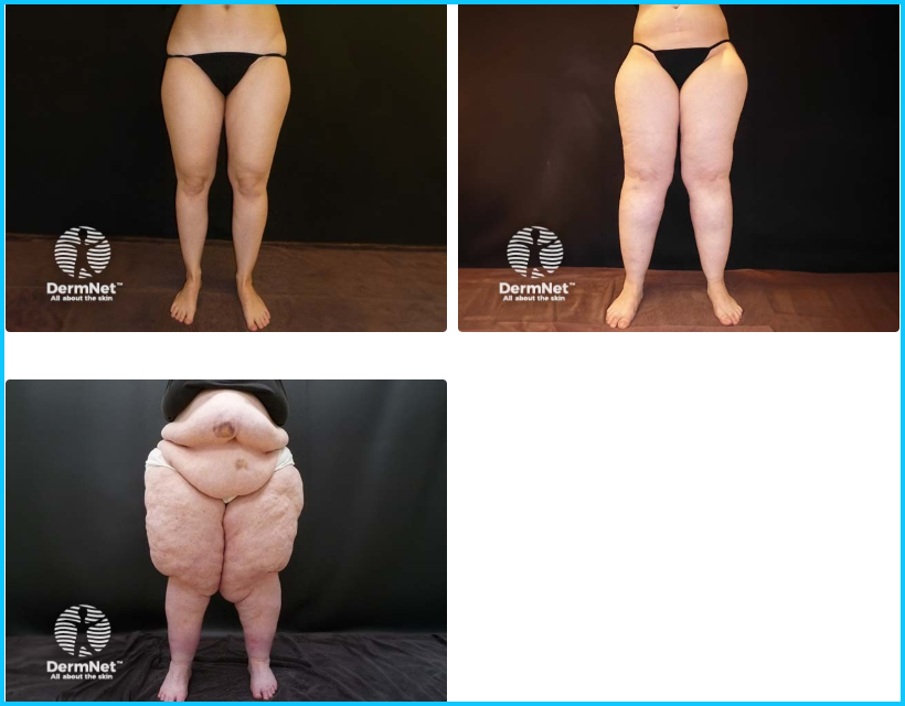
On examination, lipoedema is characterised by symmetrical, disproportionate accumulation of adipose tissue in the limbs, particularly the legs, with relative sparing of the trunk, feet and hands. Legs may appear column-shaped or lobular, with abrupt termination of fat deposition at the ankles (cuffing sign). Proliferation of fatty tissue over the medial thighs can push the legs into a valgus (knock kneed) position.
Key features that help distinguish lipedema from other chronic lower-limb changes include easy bruising, marked pain/tenderness, and nodularity of the subcutaneous tissue.
Three-stage classification (based on tissue morphology, not symptom severity):
- Stage I: Smooth skin with homogeneous subcutaneous tissue.
- Stage II: Uneven skin with palpable subcutaneous nodules.
- Stage III: Large, disfiguring fat lobules, prominent skin folds and marked nodularity.
Important: Disease stage does not necessarily correlate with pain, functional impairment or symptom severity.
Management
Lipoedema is a chronic condition that can significantly impair quality of life and function.
Management focuses on symptom relief, mobility and function.
Weight management and regular low-impact exercise are encouraged, although lipoedema-associated adipose tissue is often resistant to weight loss.
Compression garments may reduce pain, heaviness and associated oedema. Manual lymphatic drainage may provide symptomatic relief, particularly with coexisting lymphoedema, but evidence is limited.
Skin care and psychological support may also be beneficial. Specialist liposuction may be considered for selected patients with persistent, functionally limiting symptoms, although long-term evidence remains limited.
See Lipoedema-therapy (Dermnet).
Three stage classification of lipoedema
Practical management of chronic lower limb changes associated with obesity
You refer Simon to your local district nurse service for an ABI. Simon's ABI is 1.0 bilaterally.
What treatments should you offer?
Compression
Compression is the cornerstone of treatment for venous oedema and venous leg ulceration when arterial supply is adequate.
A Cochrane review found moderate-certainty evidence that compression bandages or stockings increase venous-ulcer healing compared with no compression4. Compression approximately doubled the rate of healing over time (HR 2.17, 95% CI 1.52–3.10) and increased the proportion of ulcers completely healed (RR 1.77, 95% CI 1.41–2.21). Compression probably also reduces pain.
Simply recommending compression stockings is often insufficient, particularly for people with severe obesity.
There is insufficient evidence on the most effective degree of compression. The New Zealand Expert Working Committee’s consensus is that compression should usually be within the range of 18–30 mmHg5.
Large or unusually shaped limbs, substantial oedema, reduced mobility and difficulty reaching the feet may make standard compression hosiery difficult or impossible to apply. Options may include appropriately fitted hosiery, multilayer/inelastic compression systems or adjustable compression wraps. Nursing, wound-care or lymphoedema expertise may be needed to select and fit a system that the patient can realistically use.
For established venous ulcers, in the absence of arterial disease or diabetes mellitus, graduated compression therapy aiming for > 30 mmHg (elastic) or a high-stiffness system (inelastic) is recommended. These should be applied and overseen by wound care experts in accordance with manufacturer guidelines.
Weight loss
Holistically address underlying metabolic dysfunction.
Movement
It is important to get the calf pump working.
Walking and ankle movement activate the calf muscle pump and improve venous return.
Encourage regular walking and physical activity within the person's functional capacity. For people with limited mobility, repeated ankle flexion and extension may be a realistic starting point.
Avoid prolonged periods of sitting or standing where possible. Leg elevation can help reduce dependent oedema, although it should complement rather than replace compression and movement.
A green prescription may be beneficial.
Treat the skin
Venous gravitational eczema should be actively treated rather than being mistaken for infection.
Management includes regular emollients, soap substitutes, and appropriate topical corticosteroids.
Look for fissures, interdigital maceration and tinea pedis, as disruption of the skin barrier increases the risk of cellulitis.
The mainstay of management is:
- moisturising
- treatment of venous eczema with topical corticosteroids
- appropriate wound care for ulceration or skin breakdown
- compression.
For patients with ulceration or wounds that are not responding to management in primary care, specialist wound care referral should occur depending on local district nursing guidelines.
Take regular photos to track progress. These are useful for referrals.
You arrange district nursing input to initiate appropriately fitted compression, taking into account Simon's limb size and ability to apply and remove it.
You provide education on skin care, including regular emollients, soap substitutes, treatment of active venous eczema with a topical corticosteroid, and recognising signs of skin breakdown or infection.
You discuss the contribution of obesity to venous and lymphatic dysfunction and offer a Green Prescription for supported physical activity and nutritional advice.
You encourage walking and calf-muscle exercises to improve venous return.
You arrange a review in 4–6 weeks to assess changes in oedema and skin inflammation, compression tolerance and any barriers to adherence.
Key points
- Bilateral red, swollen legs are rarely cellulitis. Consider chronic venous disease, venous eczema, lipodermatosclerosis and lymphoedema, particularly when symptoms are longstanding and antibiotics have been ineffective.
- Obesity contributes to venous hypertension and lymphatic dysfunction. These conditions frequently coexist, causing persistent oedema, inflammation and progressive skin changes.
- Recognise the characteristic skin changes. Haemosiderin pigmentation, venous eczema and lipodermatosclerosis suggest chronic venous disease. Acute lipodermatosclerosis can mimic cellulitis.
- Assess arterial perfusion before therapeutic compression. An ABI may be unreliable in patients with arterial calcification; toe pressures may be required. Compression must be appropriate to arterial status.
- Compression is the mainstay of treatment. Practical fitting, skin care, calf muscle activity, and evidence-based obesity management are important for long-term outcomes.
- Specialist nursing wound care and compression therapy is invaluable.
- Recognise red flags requiring urgent assessment:
- chronic limb-threatening ischaemia: ischaemic rest pain, non-healing wounds or gangrene
- DVT: new unilateral swelling or pain, particularly with thrombotic risk factors
- severe infection: rapidly progressive erythema, disproportionate pain, systemic toxicity or suspected necrotising infection
- bleeding varicose veins: control bleeding and arrange urgent vascular assessment.
- Recognise complications requiring referral, particularly venous ulceration and recurrent superficial thrombophlebitis, in accordance with local HealthPathways.
Content created by Dr Danuta Amelung, BHB, MbChB, FRNZCGP, AFRACMA, Dip. Paed, PGCertWHlth, PGDipTrvMed, with expert review by Dr Carl Muthu, Vascular & Transplant Surgeon.
References
Recognition of Learning Activities
Don't forget to log your time with The Royal New Zealand College of General Practitioners portal for recognition of learning activities.